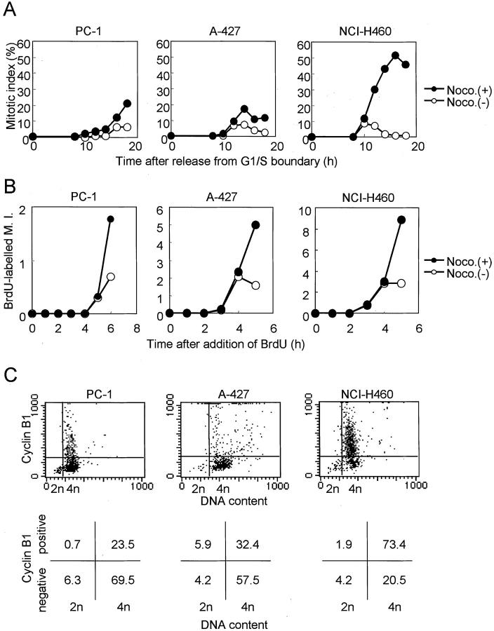
Figure 2.
Affirmation of mitotic arrest in response to nocodazole. A: Inhibition of entry into mitoses in synchronized cells. Cells that had been arrested at the S phase or G1/S boundary by 16 hours of treatment with 5 μmol/L of aphidicolin were released by replacing the culture media. The initial increases in the mitotic indices are similar regardless of the presence or absence of 200 nmol/L of nocodazole, indicating the absence of entry into mitosis in certain cell lines. B: Inhibition of entry into mitoses in BrdU-labeled asynchronous cells. Simultaneous treatment with 10 μmol/L of BrdU and 200 nmol/L of nocodazole results in the similar initial increases in mitotic cells labeled with BrdU, indicating that cell lines are not differentially arrested before entering mitosis. Note that scales of the y axes are different in each cell line. C: Flow cytometric analyses of expression of cyclin B. Upper figures represent the dot plot, and lower figures represent the percentages of cells residing in each quadrant. The majority of cells having 4n DNA content express cyclin B in a mitotic spindle checkpoint-proficient cell line, but not in the impaired cell lines.

