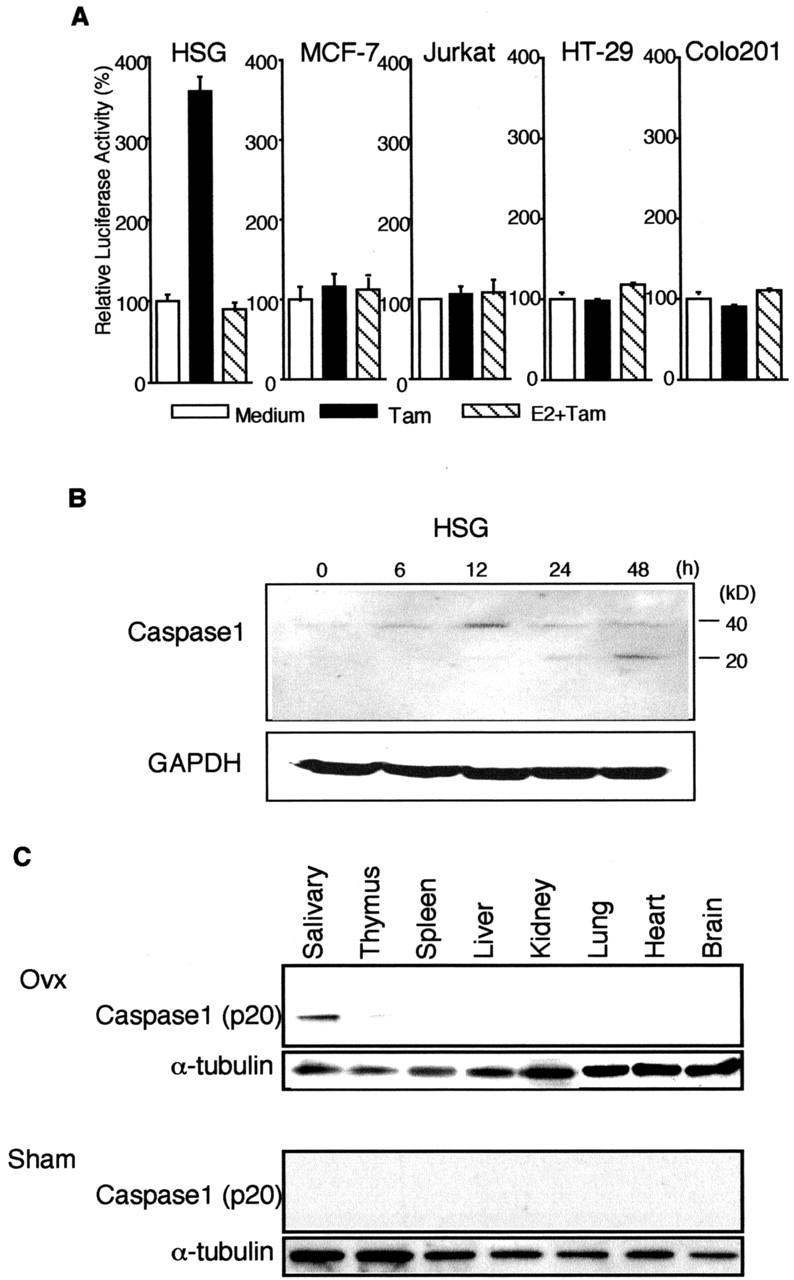
Figure 5.

A: Luciferase assay of caspase-1 promoter activities in HSG cells stimulated with Tam. HSG, MCF-7, Jurkat, HT-29, and colo201 cells, after transfection with caspase-1 promoter Luc construct, were stimulated for 2 hours without or with 1 × 10⁻⁷ M Tam. Estrogen (17β-estradiol, 1 × 10⁻⁸ M) was added to the cells 12 hours before Tam stimulation. The results are the mean values of three independent experiments run in triplicate. B: Western blot analysis of caspase-1 in apoptotic HSG cells stimulated with Tam, showing an increase in procaspase-1 (40 kd) at 12 hours, and a time-dependent increase in caspase-1 active form (p20). Cytosolic extracts were prepared from HSG cells which were treated with Tam (1 × 10⁻⁷ M) for various times. C: Detection of caspase-1 active form (p20) in the salivary gland tissue, but not in various organs from Ovx- and Sham-B6 mice on Western blot analysis. α-tubulin proteins were used as internal control. Data were representative in triplicate.
