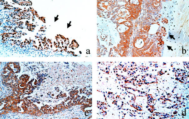
Figure 2.

Immunohistochemical staining of HIF-1α in ovarian carcinoma tissues. Nuclear localization of HIF-1α is sporadically observed in the tumor cells in the tip of the papillary projection (a) or in the vicinity of necrosis (b). Cytoplasmic localization of HIF-1α is more frequently observed (c and d). a: Serous carcinoma. b: Endometrioid carcinoma. c: Serous carcinoma. d: Clear cell carcinoma. Magnification, ×100
