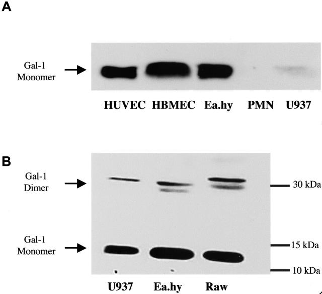
Figure 1.
Expression of Gal-1 in primary and immortalized cells. Cell extracts were subject to electrophoresis and endogenous Gal-1 expression monitored by Western blotting analysis. Extracts were prepared from U937, human umbilical vein endothelial cells (HUVECs), Ea.hy926, human bone marrow endothelial cells, human PMNs, or mouse macrophages (RAWs). A and B are representative of three distinct experiments, and show the expression of Gal-1 monomer (∼15 kd) and dimer (∼30 kd).

