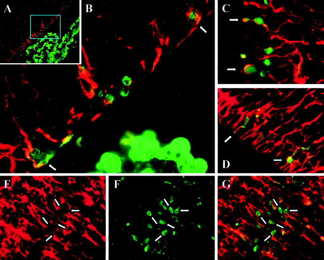
Figure 3.
Progenitor cells in the CNS are infected by CVB. Newborn pups were infected i.c. with eGFP-CVB (2 × 10⁷ pfu) and sacrificed 1 day post-infection. Paraffin-embedded transverse sections of brain tissue were deparaffinized, antigen-unmasked (see Materials and Methods), and stained to detect nestin (red) and eGFP (green). A: A low-power merged nestin/eGFP image of the lateral ventricle, and the cyan box shows the region represented at higher magnification in B. C and D: Merged images from the ventricular regions of a different neonatal mouse. E–G: Further evidence of co-localization, showing respectively nestin, eGFP, and merged images. The white arrows show examples of “empty” perinuclear areas in nestin+ cells (see text).

