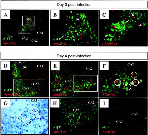
Figure 8.
Viral protein expression co-localizes with ApopTag staining within the hippocampus. Newborn pups were infected with eGFP-CVB and 3 (A–C) or 4 (D–H) days later, brains were harvested, and transverse brain sections were examined for apoptotic cells using ApopTag staining as described in Materials and Methods. Merged ×31 images (eGFP-viral protein expression; red-ApopTag) in A and D demonstrate that apoptotic areas in the hippocampus regionally localized with areas in which viral proteins were expressed. The boxed regions in A are presented at higher magnification ×62 in B and C. Nuclei which appear positive for ApopTag, and may be related to eGFP+ cells, are arrowed. Increasing magnifications of the hippocampus at 4 days p.i. (D) are shown in E ×62 and F (×100); magnified regions are boxed in the preceding panel. Cells undergoing apoptosis are circled (F), and these cells showed nuclear condensation by hematoxylin staining (G). A control section was included (H), in which the TdT enzyme was omitted during staining, thus demonstrating that the red signal is specific for apoptotic cells; and a mock-infected control brain harvested at day 4 post-injection (I) showed staining for neither eGFP, nor for apoptosis. DG, dentate gyrus; CA1, CA2, CA3, fields of Ammon’s horn.

