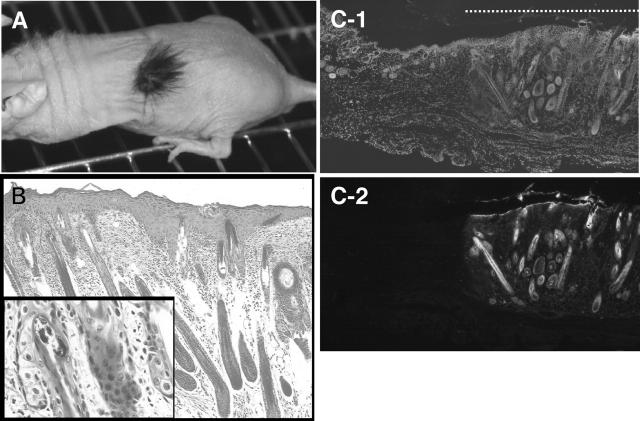
Figure 2.
Skin tissues reconstituted by a mixture of epidermal and dermal cells. A: Active hair growth in a region transplanted with embryonic epidermal and dermal cells observed 3 weeks after transplantation. B: Histology of the reconstituted skin stained with hematoxilin/eosin. Epidermis and hair follicles with sebaceous glands (inset) were seen. Bar, 50 μm (inset, 10 μm). C: Skin reconstituted by epidermal and dermal cells derived from GFP-transgenic mice. C-1: DAPI staining of nuclei. C-2: Fluorescent microscopic image. The dotted line in C-1 indicates the part of reconstituted skin. Bar, 200 μm.

