
Figure 1.
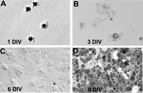
Morphologies of RPE cells at different time points of culture as indicated. Note the heavily pigmented cells at the initial stage of culture (A), the depigmented cells during subsequent culture phases (B–C), and the repigmented cells in a confluent culture (D). DIV, days in vitro. Original magnifications, ×200.