
Figure 4.
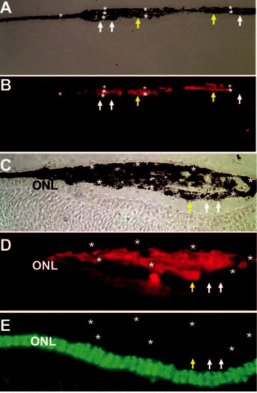
Presence of multiple layers of RPE-like tissues (stars) at the injection sites of control cells infected with RCAS-GFP and lack of visinin expression in these cells. Shown are bright-field views (A, C)or immunostaining for p27 (B, D) and visinin (E) of cross-sections of E11.5 line 72 recipient eyes. Extra tissues were composed of host cells (p27−, white arrows) and grafted cells (p27+, yellow arrows). Original magnifications: (A–B) ×200; (C–E) ×400.