
Figure 6.
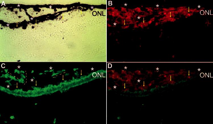
Attachment of grafted (p27+, red) and transdifferentiating (visinin+, green) cells to the ONL and rudimentary alignment of the cells along the extra RPE-like tissues. Shown is double labeling of a cross-section of E17 eye (Spafas pathogen-free embryos) for p27 (B) and visinin (C). Bright-field view (A) and simultaneous view of the double labeling (D). Dashed line: rudimentary alignment of the cells along the extra RPE-like tissues. Arrows: grafted, transdifferentiating (double-labeled) cells attached to the ONL of the host retina. Stars: RPE or RPE-like tissues. Original magnifications, ×400.