
Figure 8.
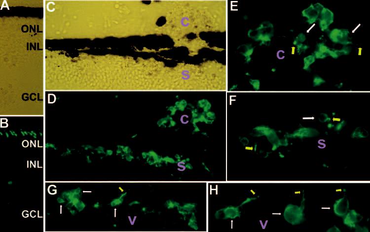
Expression of red opsin in transdifferentiating cells placed in the subretinal space (s), the choroid (c), and the vitreous (v). Shown are immunostaining of E18 recipient eyes that received grafted cells at E7.5. In E18 retina (A; bright-field view), the anti–red opsin antibody decorated the outer segments (B). At the injection site (C; bright-field view), however, such a well-formed order of the red opsin+ outer segments of host photoreceptors was not apparent (D). A large number of grafted cells were red opsin+ in the subretinal space (s in D, F), the choroid (c in D, E), and the vitreous (v in G, H). Arrows: cell bodies (white)or outer segment–like structures (yellow). Original magnifications: (A–D, G) ×400; (E, F, H) ×1000.