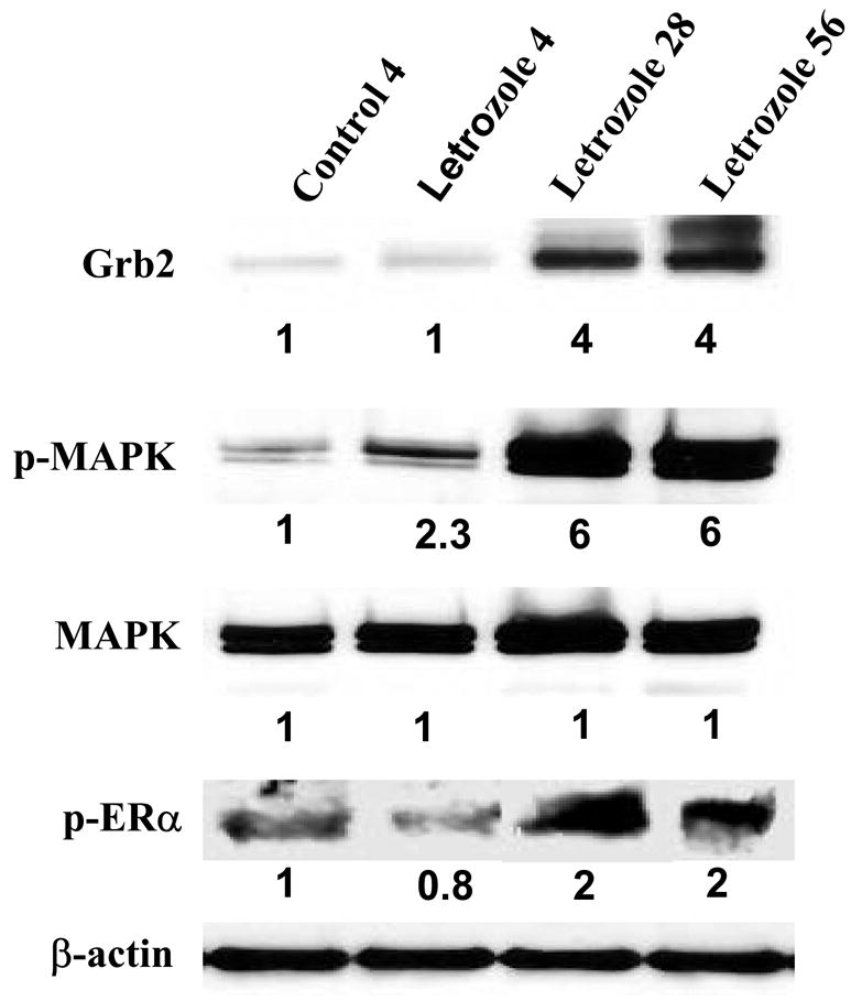
Figure 1. The Effect of Letrozole Treatment on Grb2, p-MAPK, and p-ERα¿ Expression in MCF-7Ca Tumor Xenografts.

Tumors were collected from letrozole-treated mice at 4 weeks (when they were responding to letrozole), 28 and 56 weeks (when they were growing on letrozole), analyzed by Western immunoblotting, and were compared with tumors of vehicle-treated mice collected at week 4 (control). Tumors were homogenized in lysis buffer, and equal amounts of protein (60 Ag) were separated on a denaturating polyacrylamide gel and transferred to a nitrocellulose membrane. After blocking nonspecific binding with 5% nonfat milk in PBS-T, the membranes were incubated with respective primary antibodies, and specific binding was visualized by using species-specific immunoglobulin G followed by ECL detection (ECL kit) and exposure to ECL X-ray film. After exposure to X-ray film, the membranes were stripped and probed for β-actin to confirm that equal amount of proteins were loaded in each lane. Numbers below the blots represent fold change in protein expression compared with the control obtained by densitometric analysis. (From Jelovac et al., Cancer Res., 2005).
