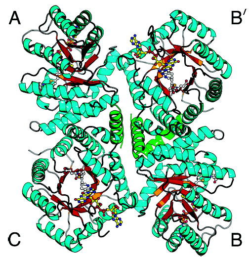
Figure 5.

The MTHFR tetramer with bound LY309887 viewed down the local 2-fold axis. As in Figure 2, sheet strands are gold and helices are cyan, with the exception of the N-terminal helix (green). The N-terminal helix, which could not be built in the initial structure, fills in the center of the tetramer. Chains are designated differently from those of our previous drawing (19).
