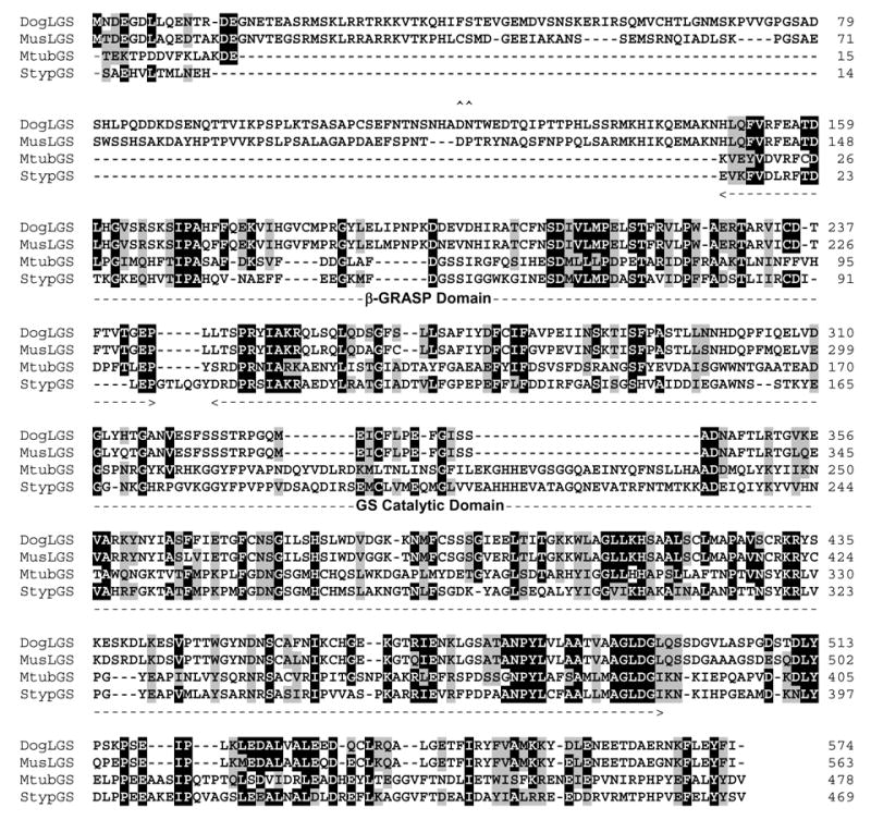
Figure 3. Comparison of lengsin and GS I sequences.

Alignment of two mammalian lengsin protein sequences (dog: ABD74632; mouse: AAN38298) with two GS I enzymes for which x-ray structures are known (Mycobacterium tuberculosis: P0A590, Salmonella typhimurium: P0A1P6). Sequences were aligned using ClustalW (with manual adjustment) in BioEdit. Conserved identical and similar residues are boxed and shaded. Extent of β-GRASP and GS catalytic domains are indicated. Bacterial sequences are numbered without the initiator methionine to correspond with the x-ray structures. the position of the Asp-Pro dipeptide that may be involved in cleavage of the N-terminal domain is indicated by ^^.
