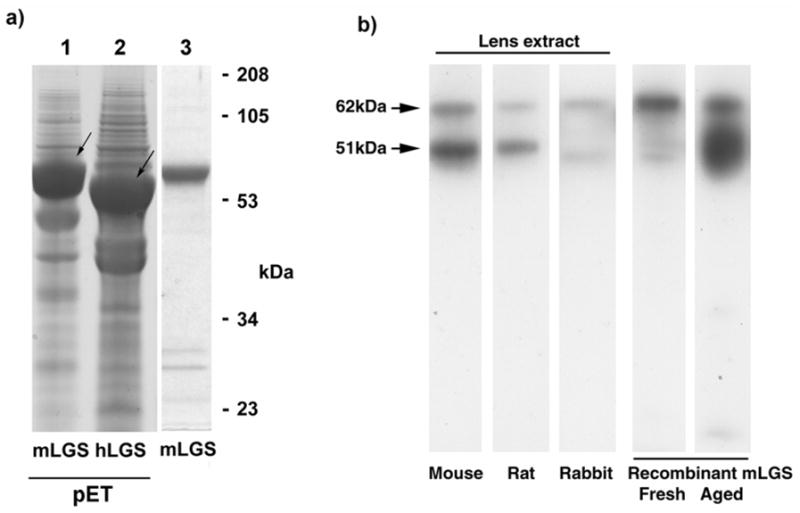
Figure 6. Recombinant protein and western blotting for lengsin.

a) Expression of recombinant human and mouse lengsin in E.coli. First two lanes show SDS PAGE of whole cell extract of cells expressing mouse or human lengsin (arrowed) in the pET system. Third lane shows purified soluble mouse lengsin (from a separate gel).
b) Western blot of mouse, rat and rabbit lens soluble extracts and aged recombinant mouse lengsin shows two bands for lengsin. The upper band corresponds in size to full length lengsin while the lower band corresponds to the size of an N-terminal truncation.
