
Figure 1. Fluorescent signal peptides label wildtype mitochondria.
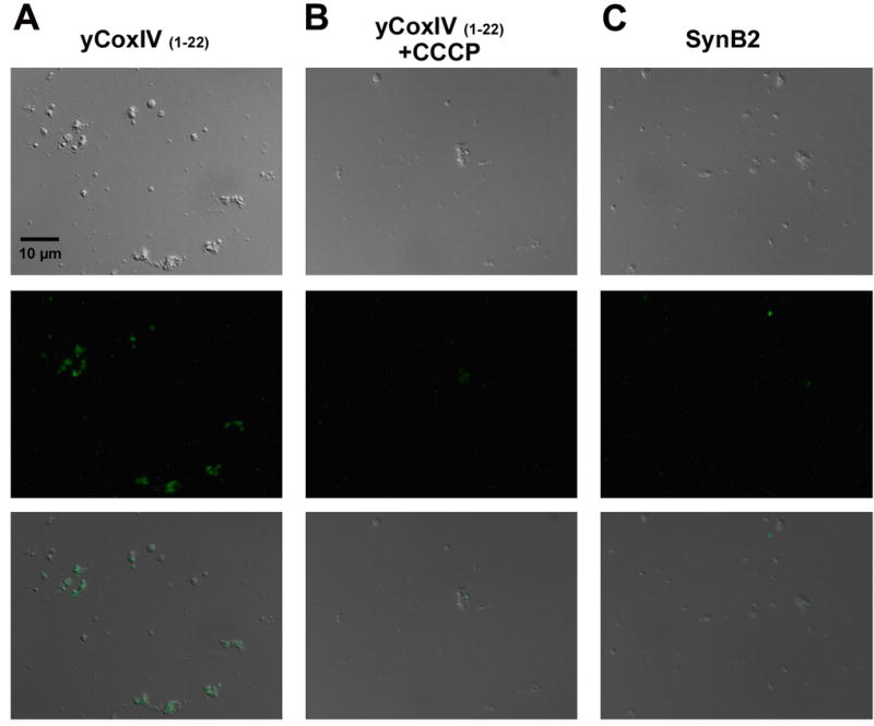
DIC (upper), fluorescence (middle) and overlay (lower) images are shown of wildtype mitochondria incubated with Alexa Fluor-yCox-IV(1-22) under energized (A) and de-energized (+1 μM CCCP) (B) conditions for 10 minutes. Images are shown of mitochondria incubated with control peptide Alexa Fluor-SynB2 under energized conditions(C). Scale bar in panel A corresponds to 10 microns for all panels.