
Figure 3. Fluorescent signal peptides label mitochondria lacking Tim44p but not Tim23p.
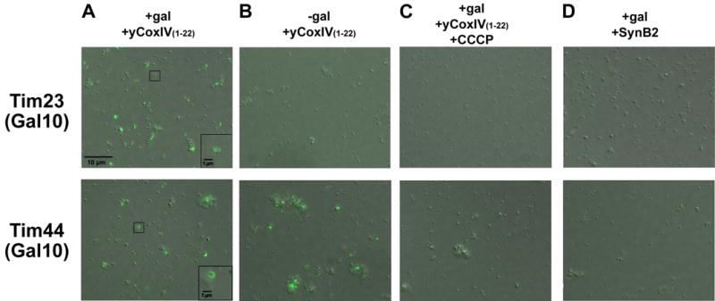
Overlays of DIC and fluorescence images are shown of mitochondria from Tim23(Gal 10) (top) and Tim44(Gal 10) (bottom) strains after a 10 minute incubation under different conditions and 3 washes to remove peptides that were not imported. Mitochondria from yeast grown with (A) or without (B) galactose were incubated with Alexa Fluor-yCox-IV1-22 under energized conditions. Mitochondria from yeast grown with galactose were incubated with Alexa Fluor-yCox-IV1-22 plus CCCP (1 μM) (C) or with the control peptide Alexa Fluor-SynB2 under energized conditions (D). Scale bar in panel A corresponds to 10 microns for all panels. Squares in lower right corner of left panels show enlarged regions with 1 micron scale bar.