
Figure 4. Electrophysiological behavior of TIM23 channels after depletion of Tim44p or Tim23p.
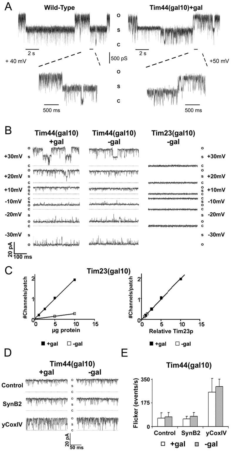
A, B, D. Typical current traces are shown of single TIM23 channels recorded from proteoliposomes containing inner membranes of mitochondria of indicated strains grown with (+gal) and without (−gal) galactose. O, S, C corresponds to the open (1000 pS), sub- (500 pS), and closed states, respectively. C. Inner membranes from Tim23(Gal 10) yeast were reconstituted with different protein concentrations. Plots show the frequency of detecting normal TIM23 channels as a function of total protein (left) and relative amount of Tim23p (right), which was determined by semi-quantitative western blots using densitometry. 12 independent patches/point are shown. D. Current traces are shown at +20 mV before (Control) and after sequential perfusion of the bath with 20 μM SynB2 and then 20 μM yCox-IV(1-13). E. Histograms of flicker rates (number of transition events/second) are shown in the absence (Control) and the presence of SynB2 or yCox-IV(1-13) for the TIM23 channels from Tim44(Gal10) yeast.