
Fig. 2.
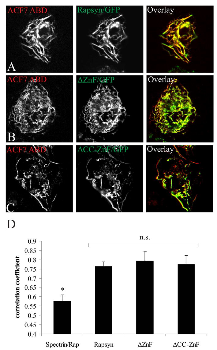
Rapsyn’s TPR domains mediate co-distribution with the ACF7 ABD in COS cells. Cells co-expressing the ABD of ACF7 and full length rapsyn/GFP (A), rapsyn ΔZnF/GFP (B) or rapsyn ΔCC-ZnF/GFP (C) were fixed and imaged as in Fig. 1. The ABD of ACF7 (red) and each rapsyn construct (green) co-distributed significantly in actin filaments (yellow in the overlay panel). (D) Quantitative analysis showed that there was no difference in co-localization with the ABD of ACF7 and each of the rapsyn constructs, but that all three constructs showed significantly higher co-distribution than the ABD of spectrin with full length rapsyn (mean ± S.E.M., n= 20 from 2–3 separate experiments). n.s., not significantly different (p > 0.1, one-way ANOVA followed by Tukey’s HSD post hoc test); *, spectrin ABD is significantly different from ACF7 ABD co-distribution with rapsyn constructs (p < 0.01); Rap, full length rapsyn.