
Figure 2.
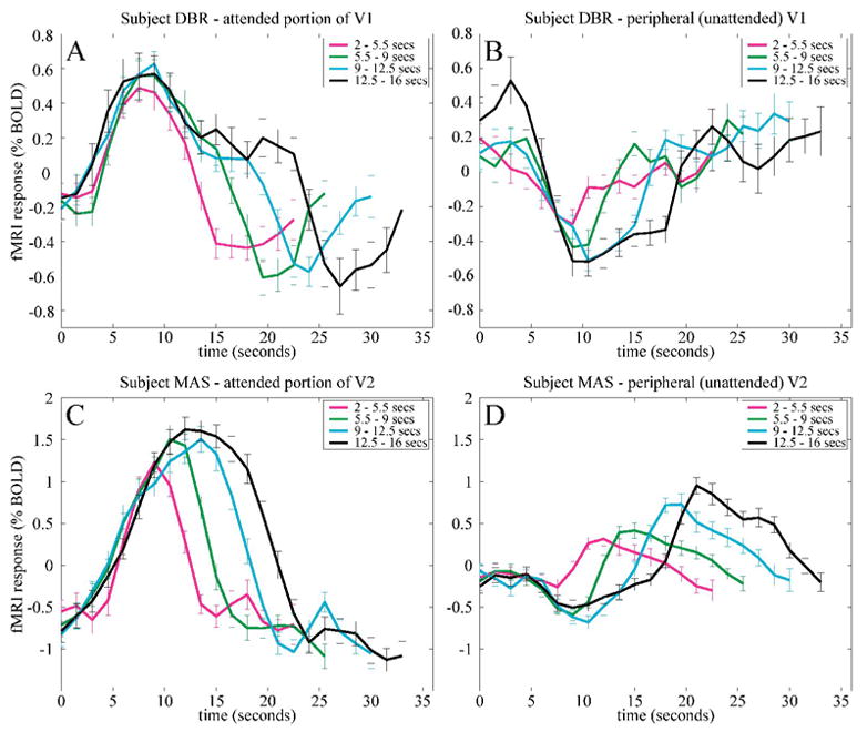
Sustained delay-period activity in early visual cortex (example data from two subjects). fMRI responses were aligned at the beginning of each trial and binned into four groups (magenta, green, cyan, black curves) based on delay-period duration. A, C. fMRI responses in a subregion of early visual cortex corresponding to the attended portion of visual field. Response increases were time-locked to the beginning of the delay period, but they returned to baseline at different times depending on the delay-period duration. B, D. Peripheral, unattended portion of visual field. Activity decreased during the delay period, but the durations of sustained decreases in activity were also a function of delay-period duration.