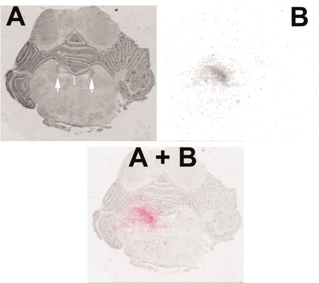
Figure 4.
A, Section from a P14 pup counterstained with cresyl violet. The white arrows mark the locus ceruleus bilaterally. Actual cannula tip placement is outside the plane of this section. B, The same section as in A at the same magnification and orientation, characterizing the extent of [3H]NE drug diffusion within the LC. A+B, Color overlay of [3H]NE diffusion on the histological section showing drug diffusion over the region of the locus ceruleus.

