
Fig. 4.
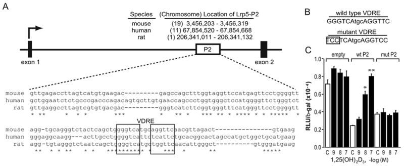
The mouse mLrp5-P2 region contains a functional VDRE. A, Conservation of P2 within the mouse, human and rat Lrp5 genes. Upper panel: The diagram depicts the boxed position of the Lrp5-P2 region located within the intron between exons 1 and 2. The nucleotide and chromosomal position of an analogous region in the human and rat genes are indicated. Lower panel: Nucleotide sequence of the P2 region with the mouse, human and rat Lrp5 gene wherein half-sites of a putative VDRE identified by the CONSITE algorithm is indicated. B, The panel illustrates the VDRE constructs used to evaluate the transcriptional activity of the potential VDRE. C, Transcriptional activity of wildtype and mutant forms of mLrp5-P2. MC3T3-E1 cells were co-transfected with pCH110-βgal, pcDNA-hVDR, and either ptk-luc control vector, ptk-mLrp5-P2 or the mutant version of ptk- mLrp5-P2. Cells were treated with either vehicle or increasing concentrations of 1,25(OH)2D3 (10−7 M) for 24 hrs and then evaluated for both luciferase and β-gal activity. Each point represents the normalized RLU average ± SEM for a triplicate set of transfections. * indicates p<0.01 compared to vehicle treated sample (one way ANOVA).