
Figure 5.
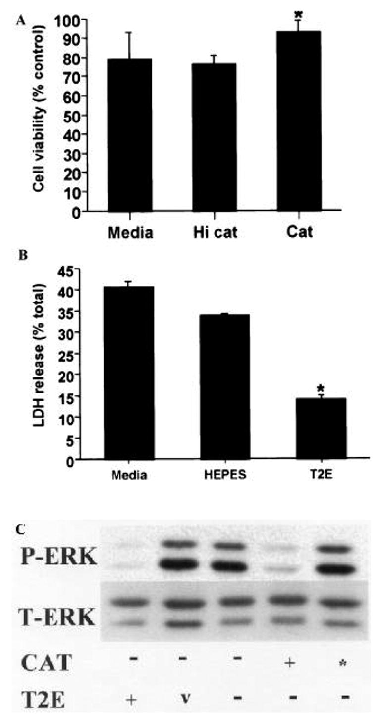
Cytotoxic doses of 6-OHDA induce ERK phosphorylation in differentiated B65 cells. Differentiated B65 cells were exposed to 500 μM 6-OHDA or vehicle for 20 h in media alone or media containing (A) catalase (Cat, 30 U/ml) or heat inactivated catalase (HI Cat), or (B) MnTE-2-PyP (T2E, 50 μM) or mimetic diluent (HEPES) and then assessed for cell injury as in figures 1 and 2, respectively. LDH release of vehicle treated cells (no 6-OHDA) was 15 ± 0.8%. *P < 0.05 by two-tailed Student’s t-test compared to 6-OHDA treated cells lacking antioxidant (Media). Representative plots are illustrated. (C) Equal protein (10 μg) from cell lysates prepared following exposure of differentiated B65 cells to 500 μM 6-OHDA for 20 h in media containing catalase (30 U/ml), heat inactivated catalase (*), MnTE-2-PyP (50 μM) or mimetic diluent (v) was subjected to immunoblot analysis for P-ERK (top) and T-ERK (bottom) as in figure 3.