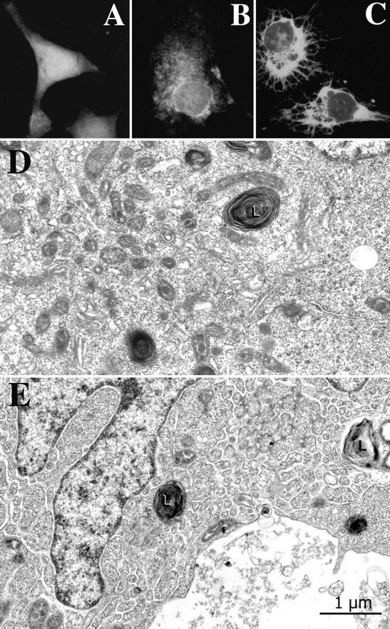
Figure 5.

Junctate-positive network in junctate overexpressing T3-HEK293 cells. (A) An example of a COS-7 cell transfected with GFP. The cell shows an amorphous cytoplasmic fluorescence. (B and C) Examples of COS-7 cells transfected with the full-length junctate-GFP. An extensive GFP-positive network extends from the perinuclear region throughout the cytoplasm. Approximately 25% junctate-GFP–positive COS-7 cells had the spindle-like appearance as shown in C. (D) GFP-transfected T3-HEK293 cells have a fairly extensive amount of smooth ER near the cell center (left portion of the image) but little of it elsewhere (at right). L, lysosome. (E) This junctate-GFP overexpressing cell exhibits a very extensive ER network, continuous with the nuclear envelope and reaching out to the cell periphery. Such an extensive network was seen in a minority of cell profiles in sections from junctate-GFP expressing cells, but never in the control cells expressing GFP. Other cells from the same cultures show variable amounts of ER.
