
Fig. 6.
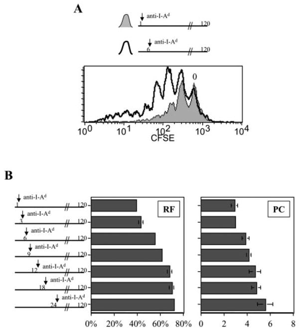
Prolonged stimulation of naive CD4+ T cells by dendritic cells influences both RF and PC. CFSE-labeled naive CD4+ DO11.10 T cells were co-cultured with pOVA (50 μg/ml)-pulsed dendritic cells for 1–24 h, after which anti-I-Ad blocking mAb was added to culture as indicated. Thereafter, T cells and dendritic cells remained in co-culture under conditions of partially inhibited TCR engagement until harvested for FCM analysis at 120 h. (A) Full antigenic stimulation for 1 h led to less T cell division than full stimulation for 6 h. The CFSE peak representing undivided cells is marked ‘0’. (B) RF and PC were calculated for the different experimental groups labeling the y axis.