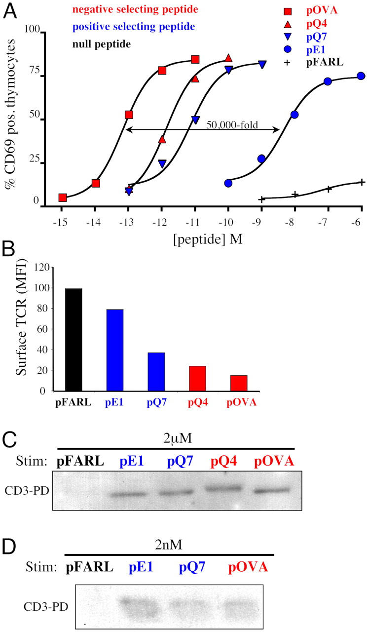
Figure 4.

Positive and negative selecting peptides induce open CD3 in thymocytes in vitro. Peptides were scored regarding their effect on thymic selection in FTOC as either null (black), positive selecting (blue), or negative selecting (red). (A) APCs loaded with varying concentrations of pFARL, pE1, pQ7, pQ4, or pOVA were cocultured with OT-I β2m−/− RAG2−/− DP thymocytes for 16 h at 37°C. Cells were then stained with anti–CD69-PE and the percentage of positive thymocytes was measured by flow cytometry. (B) APCs loaded with 2 μM pFARL, pE1, pQ7, pQ4, or pOVA were cocultured with OT-I β2m−/− RAG2−/− DP thymocytes for 30 min at 37°C. Cells were then stained with anti–TCRβ-PE and the mean fluorescence intensity (MFI) of surface TCR expression was measured by flow cytometry. (C) APCs preloaded with 2 μM pFARL, pE1, pQ7, pQ4, or pOVA were cocultured with OT-I β2m−/− RAG2−/− DP thymocytes for 30 min at 37°C. Cells were then lysed and the open CD3 PD assay was performed. (D) APCs loaded with 2 nM pFARL, pE1, pQ7, or pOVA were cocultured with thymocytes for 30 min at 37°C. The open CD3 PD assay was then performed on lysates of cells stimulated as described in C.
