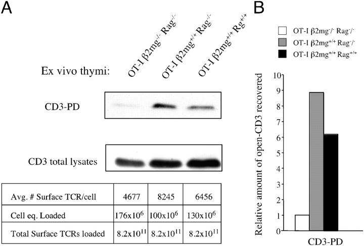
Figure 5.
The expression of cognate pMHC ligands induces open CD3 in thymocytes in vivo. (A) Thymocytes were obtained from 7-wk-old OT-I β2m−/− RAG2−/−, OT-I β2m+/+ RAG2−/−, or OT-I β2m+/+ RAG2+/+ mice. Without any exogenous stimulation, thymocytes were lysed and subjected to the open CD3 PD assay. Quantitative flow cytometry was used to estimate the average number of surface TCRs expressed on thymocytes from each genotype. These calculations were used to load each gel lane with equal numbers of surface TCR equivalents, as noted below the blot (average no. surface TCRs/cell × cell equivalents loaded = total surface TCRs loaded). For comparison, a Western blot of CD3ζ was performed on total thymocyte lysates loaded according to the same calculations. (B) Pixels from the bands obtained in A were quantified and the fold-increase in band intensity was calculated relative to the signal obtained from OT-I β2m−/− RAG2−/− thymocytes.

