
Figure 5.
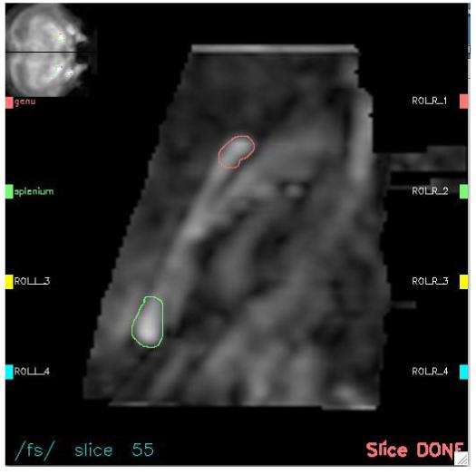
ROI identification procedure. Interactive computer interface display of an enlarged midsagittal FA image (center) with its slice location on a coronal projection (upper left hand corner). The genu (top, red) and splenium (bottom, green) have been manually delineated .