Figure 2.

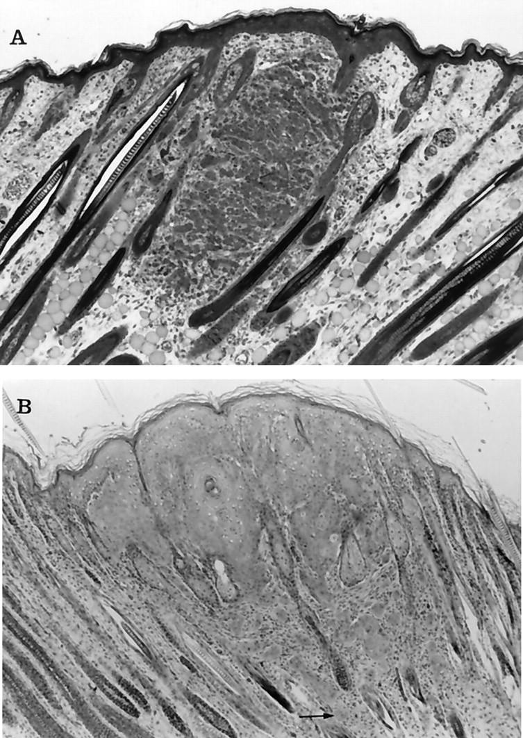
Photomicrographs of keratoacanthomas from transgenic rabbits at 1 and 3 days of age. A: At 18 hours after birth the small mass of epithelial cells appeared to be associated with hair follicles, although the overlying epidermis was also slightly thickened. The cells within the tumor were arranged in small clusters that were separated by blood vessels and connective tissue. Some of the cells contained cytoplasmic vacuoles, suggesting sebaceous differentiation. At higher power (not shown) intercellular bridges were visible between cells. B: At 3 days the tumor cells had spread laterally to replace and compress hair follicles and were contiguous with the overlying epidermis. The neoplastic cells shown squamous differentiation, including clusters of “glassy cells” and keratin horn formation. Notice the clusters of neoplastic cells that have invaded the deeper areas of the dermis separate from the main tumor mass (arrow). Toluidine blue and H&E stains, respectively. Original magnification, ×50.
