Figure 3.

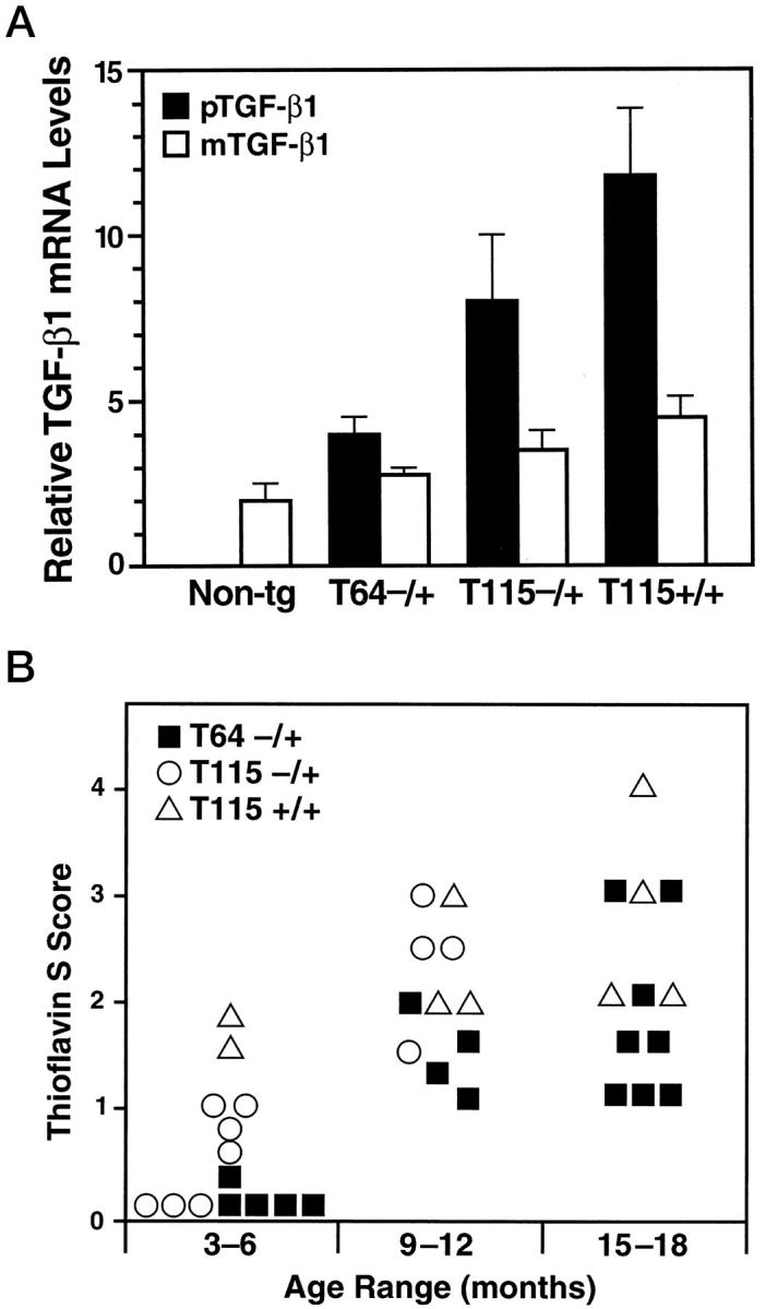
Accumulation of thioflavin S-positive deposits in TGF-β1 mice depends on age and level of TGF-β1 expression. Brains from TGF-β1 line T64 heterozygous mice (T64−/+), line T115 heterozygous mice (T115−/+), and line T115 homozygous mice (T115+/+) and nontransgenic littermate controls (Non-tg) were analyzed for cerebral TGF-β1 mRNA expression levels and for the number of thioflavin S-positive cerebrovascular deposits. A: Total RNA was extracted 3 months postnatally (n = 4 mice per group). The relative levels of porcine (transgene) TGF-β1 (pTGF-β1) and murine (endogenous) TGF-β1 (mTGF-β1) were determined by RNase protection assay. Results are means ± SEMs obtained by phosphorimager analysis and normalization to actin values. B: Sagittal brain sections from the indicated groups of mice (aged 3–6, 9–12, or 15–18 months) were stained with thioflavin S and examined by fluorescence microscopy. Data points represent mean scores from three to six sections per mouse. Older mice had more thioflavin S-positive deposits than younger mice with similar levels of transgene expression, and higher levels of TGF-β1 expression resulted in more amyloid deposits.
