
Figure 4.
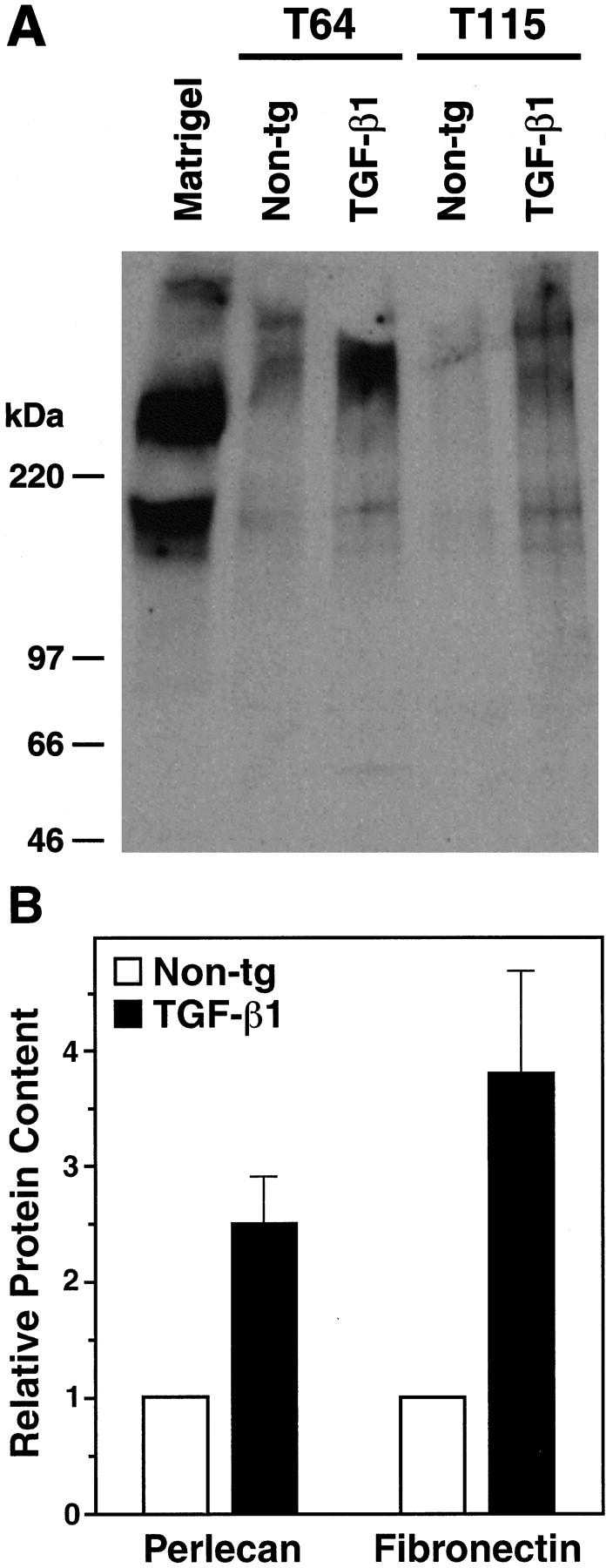
Increased perlecan and fibronectin levels in cerebral microvessels from young TGF-β1 mice. A: Western blot analysis showing perlecan expression in homogenates from isolated cortical and hippocampal microvessels (180 μg per lane) from 4-month-old TGF-β1 mice (line T64 or T115 heterozygous) or nontransgenic littermate controls. A mix of extracellular matrix proteins (Matrigel, 1 μl) served as a positive control. Microvessels prepared from TGF-β1 transgenic or nontransgenic control brains (n = 5 per group) were pooled yielding approximately 300 μg of protein per group. Molecular size standards shown on the left indicate perlecan-immunoreactive products in cerebral microvessels exeeding 400 kd, as well as a smaller-size protein of approximately 200 kd, which is also present in Matrigel. B: Perlecan and fibronectin contents determined semiquantitatively by densitometric analysis in three to four independent experiments were significantly higher in TGF-β1–transgenic (line T64) than in nontransgenic microvessels (*P < 0.05; Mann-Whitney U test). Results are means ± SEMs.