
Figure 5.
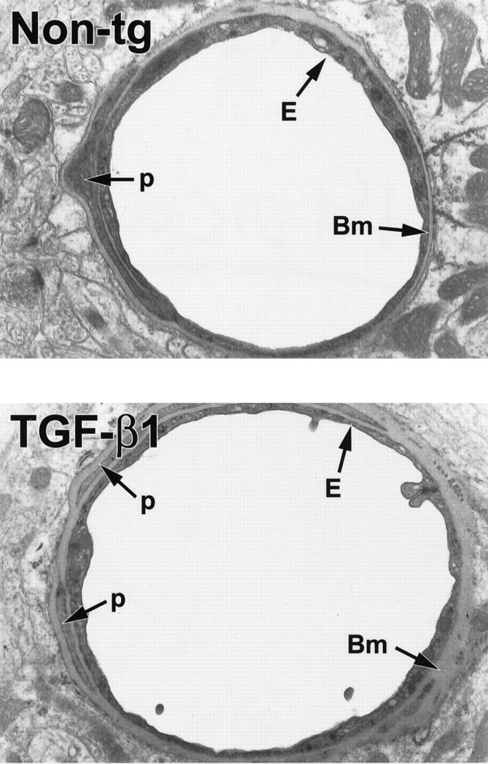
Ultrastructural abnormalities in cerebral blood vessels of TGF-β1 mice. Ultrathin brain sections from 9-month-old TGF-β1 mice (line T64 heterozygous) or nontransgenic littermate controls (Non-tg) were analyzed by electron microscopy. In the TGF-β1 transgenic vessel, the basement membrane (Bm) appears thickened, and the endothelial cell (E) profile is thinner with an irregular luminal surface. The nontransgenic control vessel has a normal basement membrane and a smooth endothelial cell surface. Original magnifications, ×5000. p, pericyte.