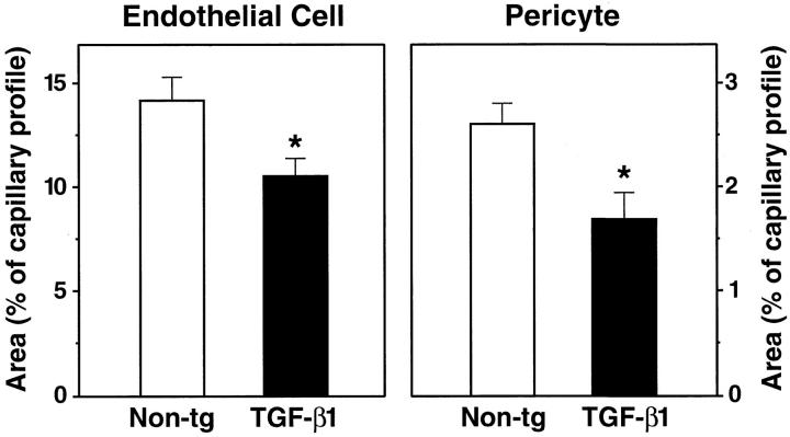
Figure 8.
Abnormalities in brain capillaries from TGF-β1 mice. The relative areas occupied by endothelial cell profiles (left) or pericyte profiles (right) were measured in cross-sections of cortical capillaries from 9-month-old TGF-β1 mice (line T64 heterozygous) and nontransgenic littermate controls. Results are means ± SDs from seven to nine capillaries analyzed in three mice per group. The relative areas of endothelial cell and pericyte profiles in 9-month-old TGF-β1 mice were significantly smaller than those in controls (*P < 0.05; unpaired, two-tailed Student’s t-test).

