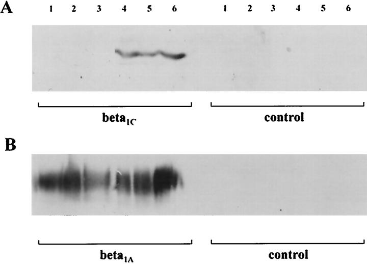
Figure 3.
Immunoblotting analysis of five breast carcinomas immunostained for β1C (A, 130–140 kd) and β1A (B), as described in the text. B: Normal expression of β1A in all of the samples. Lanes 1–3: The three cases that were not immunoreactive for β1C did not show any immunohistochemical staining as well. Lanes 4 and 5: The two immunoreactive cases were also intensely decorated by the antibody in tissue sections. Lane 6: Positive control (normal prostate).

