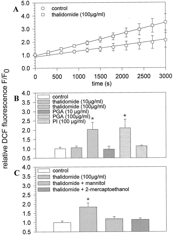
Figure 2.
Generation of hydroxyl radicals by thalidomide. Embryoid bodies were preincubated with thalidomide for 4 hours. Subsequently they were stained with the ROS indicator H2DCF-DA and the generation of fluorescent DCF was monitored. A: Time course of ROS generation in control and thalidomide-treated embryoid bodies after a 4-hour incubation with the compound. Data were fitted by linear regression. B: ROS generation by different concentrations (10 μg/ml and 100 μg/ml) thalidomide, PGA, and PI (100 μg/ml). C: Effects of the hydroxyl radical scavengers mannitol (10 mmol/L) and 2-mercaptoethanol (300 μmol/L) on ROS generated by thalidomide in embryoid bodies. Control DCF fluorescence was set to 100%. Significance (P < 0.05) is indicated by an asterisk.

