
Figure 4.
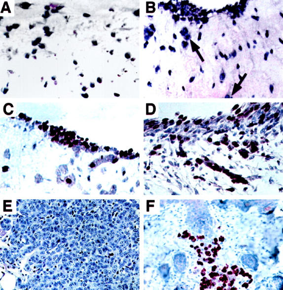
Survival, growth, and migration of RGP melanoma cells Sbcl2 in dermal equivalent after transduction with bFGF gene. A: Sbcl2 melanoma cells transduced with lacZ control gene and embedded together with fibroblasts in dermis were stained with hematoxylin and eosin. Magnification, ×50. B: Sbcl2 cells transduced with bFGF and mixed with fibroblasts in collagen form small clusters in the collagen and migrate out of the matrix for nest formation (arrows). Magnification, ×50. C: Sbcl2 cells transduced with LacZ control vector and stained for proliferation marker Ki67. Magnification, ×50. D: Sbcl2 cells transduced with bFGF, stained for Ki67. Section was counterstained with Mayer’s hematoxylin. Magnification, ×50. E: H & E stain of tumor from SCID mouse injected s.c. 4 days earlier with bFGF-transduced Sbcl2 cells of section. Magnification, ×40. F: Section of bFGF-transduced Sbcl2 cells stained after 14 days for Ki67 proliferation marker. Magnification, ×40.