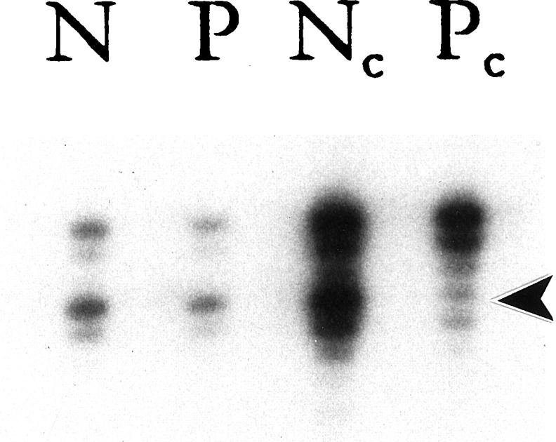
Figure 2.
Representative example of X chromosome inactivation at the androgen receptor locus. DNA samples from normal tissues (N) and papilloma (P) were subjected to a mock reaction (rows 1 and 2) or digested with HhaI (rows 3 and 4, c = cut) followed by PCR amplification. After digestion with HhaI, the normal specimen revealed no change in relative intensity of the two alleles, a normal polyclonal pattern. Conversely, the papilloma displayed a significant reduction in the intensity of the lower allele (arrowhead) as compared to the upper allele, a typical monoclonal pattern.

