
Figure 1.
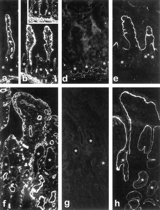
Expression and distribution of laminins in the small intestine from patients with CD. Indirect immunofluorescence micrographs from representative fields of cryosections from control specimens (a) as well as uninflamed (b–e) and inflamed (f–h) paired regions of CD specimens stained for the detection of laminin α1 (a–c, f), α2 (d, g), and α3 (e, h) chains. The α1 chain was found to be expressed in the basement membrane of villus cells under all conditions (a–c, f) as well as in crypts (asterisks) of uninflamed (b, c) and inflamed (f) CD specimens, but not in crypts of control specimens (a). The α2 chain remained normally expressed in uninflamed CD specimens, being restricted to the lower half of the crypts (d), whereas it was consistently undetectable in most inflamed CD specimens (g). The α3 chain was detected only in the basement membrane of the villus in uninflamed tissues (e), whereas it was found to stain both villus and crypt in their inflamed counterparts (h). Original magnification, ×128. Asterisks denote crypts.