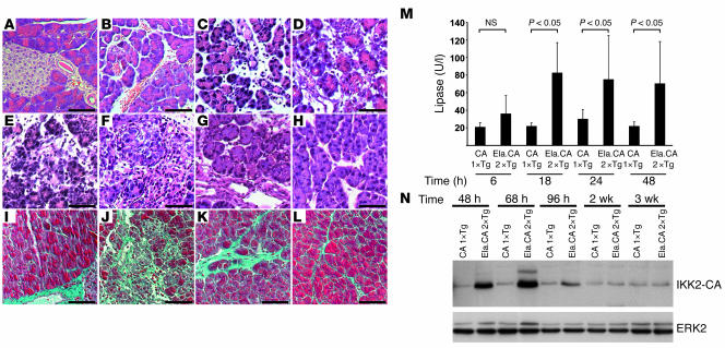
Figure 4. Overexpression of IKK2-CA induces pancreatitis.
(A–H) Histological evaluation of pancreatic sections after Dox injection showed normal morphology in controls (A, 36 hours after Dox) and pancreatitis in Ela.rtTA×IKK2-CA mice (B–H). Early signs (B; 6 hours) included intralobular edema, followed by infiltration of inflammatory cells (C; 18 hours) and acinar cell necrosis after 48 hours (D). Extended acinar cell necrosis and intralobular deposits of extracellular matrix were evident 68 hours (E) and 96 hours (F) after Dox injection. Tissue regeneration was obvious 14 days (G) and 21 days (H) after Dox injection. (I–L) Masson-Goldner connective tissue staining of the control (I), 96 hours (J), 14 days (K), and 21 days (L) after Dox injection. (M) Serum lipase was increased in Ela.rtTA×IKK2-CA mice 6 to 48 hours after Dox injection. (N) Immunoblot confirmed prolonged IKK2-CA expression in Dox-induced Ela.rtTA×IKK2-CA mice up to 96 hours after Dox injection. ERK2 expression serves as loading control. Scale bars: 50 μm.

