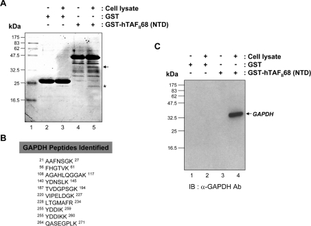
Figure 1. Identification of GAPDH as an hTAFII68-TEC-associated protein.
(A) SDS/PAGE of hTAFII68 (NTD) complexes isolated from HEK-293T cells. Total HEK-293T cell lysates were incubated with either GST or a GST-fusion protein containing the NTD of hTAFII68 (indicated above the panel). Complexes were resolved by SDS/PAGE (15% gels) and stained with Coomassie Blue. Molecular mass markers are shown on the left; they are derived from prestained protein standards (broad range; New England Biolabs). The band investigated in this analysis is indicated by the arrow to the right. Lane 1, molecular mass marker; lane 2, GST; lane 3, GST plus cell lysate; lane 4, GST–hTAFII68 (NTD); lane 5, GST–hTAFII68 (NTD) plus cell lysate. (B) Peptide sequences of GAPDH identified by MALDI-TOF analysis. The GAPDH peptides matched by MALDI-TOF are shown with amino acid numbers displayed at both ends. (C) Association of hTAFII68 (NTD) with GAPDH. HEK-293T cell lysate was incubated with either GST–hTAFII68 (NTD) or GST. After affinity-selection, the pellets from GST pull-down assays were analysed by SDS/PAGE (15% gels), and bound GAPDH was detected with anti-GAPDH antibody (MAB374; Chemicon) and chemiluminescence (Perkin Elmer Life Science). The positions of the molecular mass markers are indicated on the left-hand side, and the GAPDH band is indicated by the arrow on the right-hand side. Lane 1, GST; lane 2, GST plus cell lysate; lane 3, GST–hTAFII68 (NTD); lane 4, GST–hTAFII68 (NTD) plus cell lysate. IB, immunoblotting; Ab, antibody.

