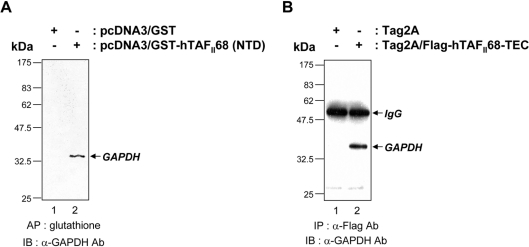
Figure 3. Association of hTAFII68-TEC with GAPDH in vivo.
(A) Co-affinity purification of hTAFII68 (NTD) with GAPDH from COS-7 cells. After transfection (48 h) of COS-7 cells with 10 μg of either pcDNA3/GST or pcDNA3/GST–hTAFII68 (NTD), cell extracts were prepared as described in the Materials and methods section and affinity-precipitated with glutathione–Sepharose beads. After fractionation by SDS/PAGE (15% gels), the proteins were analysed by Western blot with an anti-GAPDH antibody (MAB374). The identities of the transfected DNAs are indicated above the panel. The positions of the molecular mass markers are indicated on the left-hand side and the position of GAPDH is indicated by the arrow on the right-hand side. Three independent experiments were performed, all of which gave similar results. AP, affinity precipitation; IB, immunoblotting; Ab, antibody. (B) Co-immunoprecipitation of hTAFII68-TEC and GAPDH in vivo. After transfection (48 h) of HeLa cells with 10 μg of either Tag2A or Tag2A/Flag-hTAFII68-TEC, cell extracts were immunoprecipitated with an anti-Flag antibody (M2), resolved by SDS/PAGE (15% gels), and probed with an anti-GAPDH antibody (MAB374). The identities of the transfected DNAs are indicated above the panel. The positions of the molecular mass markers are indicated on the left-hand side, and the positions of IgG and GAPDH are indicated by arrows on the right-hand side. Three independent experiments were performed, all of which gave similar results. IP, immunoprecipitation; IB, immunoblotting; Ab, antibody.

