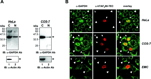
Figure 6. Co-localization of hTAFII68-TEC and GAPDH.
(A) Subcellular localization of GAPDH in HeLa and COS-7 cells. To determine the subcellular location of GAPDH, HeLa and COS-7 cells were fractionated into nuclear and cytoplasmic preparations. Proteins were separated by SDS/PAGE (15% gels) and detected by Western blotting using anti-GAPDH (MAB 374, upper panels) or anti-actin (I-19, lower panels) antibodies. Three independent experiments were performed, all of which gave similar results. IB, immunoblotting; Ab, antibody. (B) Subcellular distribution of hTAFII68-TEC and GAPDH. HeLa, COS-7, and human chondrocyte cells (C28/I2) grown on coverslips were transfected with a mammalian expression vector encoding Flag-tagged hTAFII68-TEC protein. The transiently transfected cells were fixed with an acetone/methanol mixture and incubated with primary antibodies for GAPDH (V-18) or Flag tag (M2). The subcellular distribution of GAPDH or hTAFII68-TEC was examined using a confocal laser scanning microscope (LSM5 Pascal, Carl Zeiss Co., Ltd.). The merged image (overlay) shows the co-localization. Three independent experiments were performed, all of which gave similar results.

