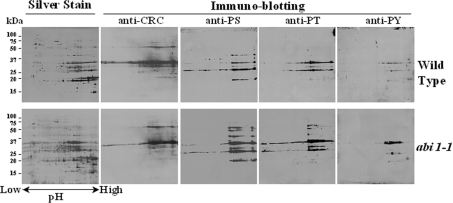
Figure 3. 2-DE gel electrophoresis and immunoblotting of seed proteins from A. thaliana cv. Lansberg erecta wild-type or the abi1-1 mutant.
Extracted proteins (1 mg) were separated by 2-DE and visualized with silver stain (indicated) or subjected to immunoblotting with anti-cruciferin antiserum (anti-CRC) or anti-phosphoserine (anti-PS), anti-phosphothreonine (anti-PT) and anti-phosphotyrosine (anti-PY) monoclonal antibodies. Molecular-mass markers (kDa) are shown in the left hand margin.

