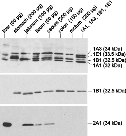
Figure 1. Immunodetection of SULTs in various parts of the gastrointestinal tract using antisera raised against rat SULT1E1 (top panel), human SULT1B1 (middle panel) and human SULT2A1 (bottom panel).
Cytosolic fractions were prepared from hepatic and gastrointestinal tissues. The amount of cytosolic protein used is indicated in parentheses. Cytosolic protein of Salmonella Typhimurium TA1538-SULT1E1 (0.5 μg) and inclusion bodies of SULT1A1 (60 ng), SULT1A3 (60 ng) and SULT1B1 (80 ng) were mixed (lane headed ‘1A1, 1A3, 1B1, 1E1’) to imitate their expression in ileum. The samples were electrophoresed and transferred to a nitrocellulose membrane. The blot was first probed with anti-r1E1-serum. After removing the antibody, the blot was reprobed with anti-1B1-serum. Antibodies were again removed and the blot was reprobed with anti-2A1-serum. Native antisera were used in these experiments.

