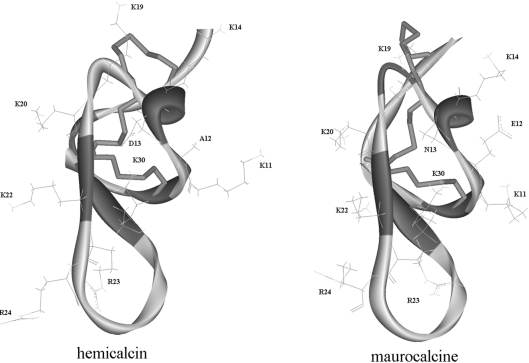
Figure 6. Homology model of HCa.
Backbone ribbon representation of the model of HCa and comparison with the structure of MCa (PDB code 1C6W [11]), which, besides IpTx A, is one of the two structures used as template for molecular modelling. Disulfide bridges are in stick representation. Basic amino acid residues present on the same face of MCa and HCa (Lys19, Lys20, Lys22, Arg23, Arg24 and Lys30) are shown. The side-chain bonds of Lys11, Lys14, Asp13 and Ala12 in HCa and Lys11, Lys14, Asn13 and Glu12 in MCa are also shown.

