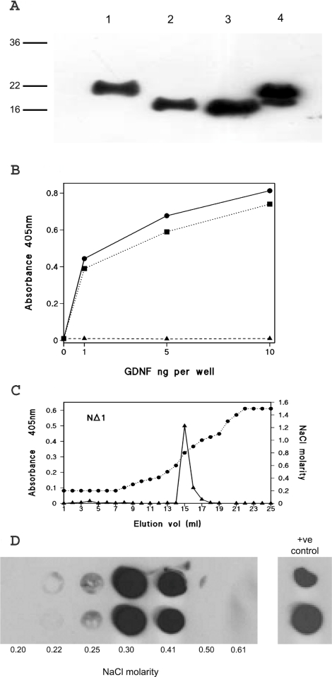
Figure 5. Characterization of partial N-terminal deleted mutants of GDNF.
(A) Western blot of baculoviral-expressed mutants of GDNF following SP-Trisacryl ion-exchange chromatography. Lane 1, wild-type rat GDNF; lane 2, NΔ1-GDNF; lane 3, NΔ2-GDNF; and, lane 4, commercial mammalian cell expressed rat GDNF. (B) Heparin-binding ELISA of wild-type rat GDNF (closed circles and solid line), NΔ1 (closed squares and dotted line), and NΔ2 (closed triangles and dashed line). Each data point is the mean of triplicate wells. (C) and (D) Heparin affinity chromatography of GDNF N-terminal deletion mutants. (C) NΔ1; immunoreactivity detected by heparin-binding ELISA. (D) NΔ2; immunoreactivity detected by dot blotting. Duplicate dot blots of fractions from the beginning of the salt gradient are shown together with the molarity determined, this region of the elution being the only one showing immunoreactivity. Duplicate applications of the input are shown on the right-hand side.

