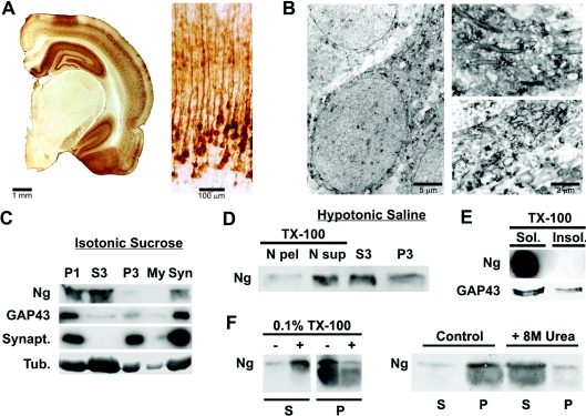
Figure 1. Ng is associated to neuronal membranes in rat brain.
Brains from young adult Wistar rats were processed for immunocytochemistry as described in the Experimental section. (A) Left: stereomicroscope print showing half a coronal section displaying abundant Ng staining at the hippocampus, cerebral cortex and amygdala and none in the thalamus. Right: bright-field print showing Ng immunostaining distribution across somatosensory cortical layers. (B) Electron microscopy prints showing typical Ng immunostaining of cortical pyramidal neurons. Ng is distributed as small aggregates along an apical dendrite and frequently labels the cytoplasmic face of intracellular membranes. (C) Brains were density-fractionated in isotonic sucrose and processed by Western blotting using specific antibodies. Synapt., synaptophysin; Tub., tubulin. P1, S3, P3, My and Syn are described in the Experimental section. (D) Brains were density-fractionated in hypotonic saline and processed by Western blotting. Most of the Ng present at the P1 fraction was recovered in a soluble form after Triton X-100 (TX-100) extraction. N pel, N sup, S3 and P3 are described in the Experimental section. (E) Triton X-100 (TX-100)-insoluble (Insol.) (lipid rafts) and -soluble (Sol.) fractions were analysed by Western blotting. No Ng could be detected in the lipid raft fraction, whereas GAP-43 was clearly partitioned between Triton X-100-soluble and -insoluble fractions. (F) Synaptosomes from sucrose density fractionation were washed with isotonic sucrose and homogenized in 20 ml of a solution containing 1 mM EDTA and 10 mM Mops, pH 7.4. After stirring for 10 min at 4 °C, the mixture was centrifuged at 15000 g for 10 min at 4 °C and the pellet was resuspended and frozen in the same buffer at 1.5 mg/ml total protein. For assays, aliquots of synaptosomal membranes were thawed, centrifuged, resuspended in the same buffer with different additions, incubated for 30 min at 4 °C with gentle stirring and centrifuged again. Aliquots of supernatant (S) and pellet (P) were analysed by Western blotting. TX-100, Triton X-100.

