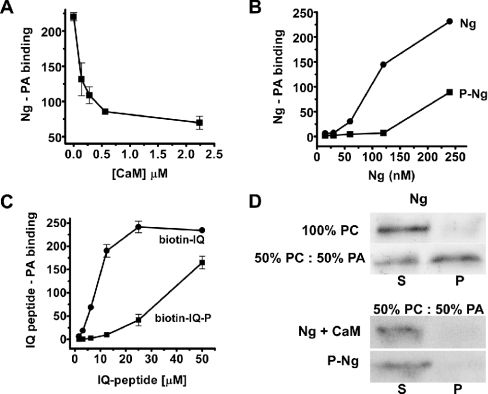
Figure 3. Regulation of Ng binding to PA.
(A) Ng binding to PA was analysed in protein–lipid overlays as described in the text, except that Ng was incubated previously with different amounts of CaM for 30 min at 25 °C before the mixture was added to egg-yolk PA-spotted strips. (B) Recombinant Ng was phosphorylated by PKC and separated from non-phosphorylated Ng by affinity chromatography in CaM–agarose columns. Binding of Ng (●) and phospho-Ng (■) was assayed as described in the text. (C) Binding of biotin–IQ peptide (●), corresponding to Ng-(29–47), and its phosphorylated form (biotin–IQ-P) (■) to egg-yolk PA-spotted strips was analysed using the method described for protein–lipid overlay assays, except that the primary and secondary antibodies were replaced by an incubation with HRP-labelled streptavidin (1:2000 dilution) for 1 h. Results in (A), (B) and (C) are means±S.E.M. for at least three independent experiments. (D) To analyse CaM competition, 5 μg of CaM was added to liposomes of the indicated composition along with Ng to the binding mixture. Typical Western blots representative of three independent experiments are shown. S, supernatant; P, pellet.

