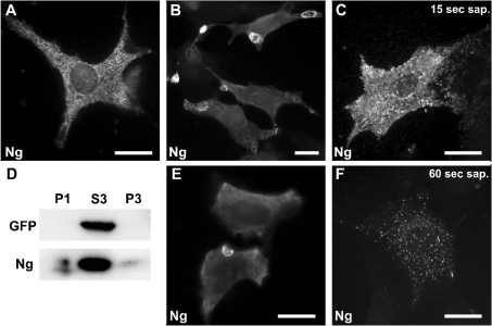
Figure 4. Ng subcellular distribution in NIH-3T3 cells.
NIH-3T3 cells were transfected with Ng alone or in conjunction with GFP. (A) Typical distribution of Ng in cells processed using the conventional IF method, showing a characteristic punctate intracellular labelling. (B, E) Ng distribution in cells processed using the cool IF method. Ng accumulates at ring-shaped structures that appear to form at the cell periphery. (C, F) Ng-transfected cells were permeabilized in situ with 0.2% saponin (sap.) for 15 s (C) or 60 s (F) and then processed using the cool IF method. Note the clustered distribution of Ng-labelled granules that spread out from the cell after 15 s of permeabilization and the spotty vesicular distribution observed after 60 s. Scale bar, 25 μm. (D) Cellular extracts from Ng- and GFP-transfected cells were density-fractionated and analysed by Western blotting. Note that, while GFP was completely recovered in the S3 fraction, visible amounts of Ng were present in the P1 and P3 fractions. P1, S3 and P3 are described in the Experimental section.

