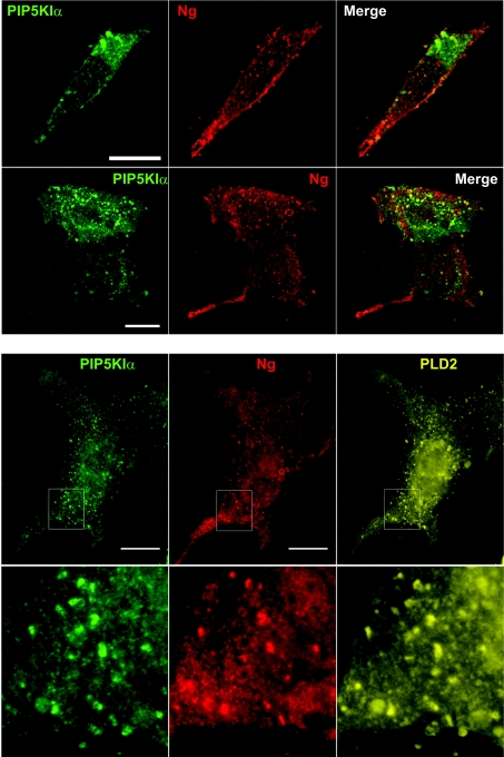
Figure 7. Effect of PtdIns(4,5)P2 availability on Ng subcellular distribution.
Upper panels: NIH-3T3 cells were transfected with Ng and PIP5KIα and their distribution was analysed using the cool IF method. In both motile (upper row) and quiescent (lower row) cells, Ng showed a predominant distribution at the plasma membrane, characterized by its presence in multiple small protrusions or blebs that displayed some co-localization with PIP5KIα. Lower panels: NIH-3T3 cells were transfected with Ng, PIP5KIα and PLD2–HA (haemagglutinin) and their distribution was analysed as described above. The expression of PIP5KIα also alters PLD2–HA localization at the plasma membrane. PIP5KIα, PLD2–HA and Ng show a typical spotty distribution, characterized by their presence in multiple small protrusions or blebs at the cell periphery. Outlines in the upper row are shown magnified in the lower row. Co-localization of P5KIα, PLD2–HA and Ng can be observed at the protrusions.

